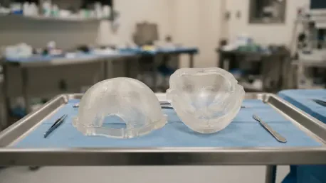

Relying on a single-sided concave mold often produces an uneven and excessively thick implant that obstructs proper anatomical alignment during intraoperative fitting. This specific challenge highlights a critical bottleneck in the democratization of 3D-printed medical devices within the neurosurgical landscape of 2026. While the ability to fabricate patient-specific implants has shifted from specialized industrial labs to hospital-based manufacturing units, the transition has faced significant technical hurdles. The promise of customized cranial reconstruction lies in its ability to restore both neurological protection and aesthetic symmetry, yet success remains contingent on a meticulous workflow that bridges the gap between digital precision and surgical reality. As medical practitioners increasingly integrate additive manufacturing into their standard of care, standardized protocols become paramount to avoiding common pitfalls that can lead to intraoperative delays or suboptimal patient outcomes. Understanding these technical nuances is no longer just for biomedical engineers; it is now a vital competency for the modern neurosurgical team.
1. Capturing Precise Radiological Data and Patient Privacy:
The production trajectory for any successful cranioplasty begins with radiological data acquisition, where the physical precision of the finished mold depends directly on the quality of the input scans. While lower-quality scans can theoretically be employed in emergency scenarios, the established standard for 2026 involves thin-slice computed tomography (CT) scans. These scans feature slice thicknesses of under one millimeter, which collectively generate approximately 150 individual spatial layers. This high level of resolution provides the necessary spatial data for accurate computer modeling, ensuring that the final print reflects the minute contours of the patient’s skull. Without this granularity, the resulting mold may fail to account for subtle bone thickness variations, leading to an implant that fits poorly within the defect. Consequently, clinical teams must prioritize these high-resolution imaging protocols to establish a reliable foundation for all subsequent manufacturing steps.
To safeguard patient privacy while maintaining procedural accountability across all manufacturing phases, patient CT data are systematically anonymized before transmission to the modeling team. Each file is assigned a unique tracking number, removing identifiable personal information while preserving the link to the clinical case. This tracking protocol is essential because it allows technicians and surgeons to trace every processing stage back to the original source data if unexpected flaws arise during printing or surgical placement. In the event of a material failure or an anatomical misalignment, the unique tracking number ensures that the root cause can be identified without compromising data security. This dual focus on data quality and administrative security creates a robust framework that supports both the technical requirements of the engineering team and the ethical obligations of the medical institution.
2. Constructing Digital Blueprints Through Collaborative Design:
Digital modeling reconstructs missing cranial anatomy by mirroring the patient’s healthy contralateral skull structure, aiming to produce a shape that matches the defect margin. However, designers must account for the fact that surgical defect borders are rarely cut perfectly perpendicular around their circumference. Cranial defects routinely exhibit a conical shape where the outer pericranial opening is wider than the endocranial margin. To address this, digital models are designed to produce an implant based on the wider pericranial edges rather than the inner contours. Furthermore, these models typically target an implant thickness of approximately 3.5 millimeters rather than attempting to match the full, variable depth of the skull. This design choice accelerates the intraoperative fitting process and reduces the volume of material required, ensuring the final product remains lightweight and biocompatible without sacrificing structural integrity.
Because unsupervised automation across digital modeling remains difficult to achieve, a joint verification step is mandatory before committing to the physical printing process. Unattended software algorithms can occasionally generate anatomically incorrect mold geometries that do not account for real-world surgical constraints. To mitigate this risk, the attending neurosurgeon and the biomedical engineer must formally review and approve the digital models. This collaboration allows the team to anticipate intraoperative complications, such as potential interference with dural structures or difficulty in securing fixation plates. By identifying these issues in the virtual environment, the clinical team avoids the high costs and significant time delays associated with printing an unusable mold. This verification protocol ensures that the physical object delivered to the operating room has been vetted for both anatomical accuracy and surgical feasibility.
3. Navigating Material Selection and Printing Realities:
The physical production of molds involves executing complex print cycles that are prone to unforeseen interruptions. In many clinical settings, 3D printing may stop prematurely due to surface impurities on the build platform or non-optimal machine calibration settings. These incomplete print cycles disrupt manufacturing workflows and generate significant material waste, leading to schedule delays that can postpone planned surgeries. To manage these risks, clinical teams are advised to schedule operative interventions only after the physical molds have been successfully fabricated and verified. Relying on a theoretical production timeline is dangerous, as the unpredictable nature of additive manufacturing can easily derail a surgical schedule. Maintaining a buffer between the completion of the mold and the start of the procedure is a fundamental logistical requirement for any hospital-based 3D printing unit.
Selecting suitable printing materials requires a careful balance between equipment costs, supply availability, and physical performance. Through clinical practice, it has been determined that some mold pliability is distinctly preferable to complete structural rigidity, as flexible molds facilitate the mechanical demolding of the cured acrylic. Surgeons and engineers must also monitor regional distribution channels to avoid disruptions in filament procurement, which can stall production for weeks. Beyond flexibility, the chosen materials must be chemically inert to prevent any adverse reactions with the bone cement during the casting process. Some low-quality filaments can create a hairy surface texture during printing, leaving micro-fibers that may adhere to the finished implant. Therefore, maintaining a reliable stock of high-quality, pliable filaments is a prerequisite for ensuring that the reconstruction process remains safe and efficient.
4. Implementing Sterilization and Intraoperative Casting Timing:
Material sterilizability represents a crucial parameter in the manufacturing workflow, as unsuitable techniques can cause structural deformation. While traditional autoclaving uses high heat that can warp many 3D-printed polymers, cold plasma sterilization has emerged as the preferred processing approach. This method ensures reliable surgical sterilization without inducing morphological distortion across the printed mold geometries, maintaining the precision established during the digital design phase. If a mold loses its shape during sterilization, the resulting implant will not fit the patient, rendering the entire preparatory process useless. Ensuring that the hospital’s sterilization department is equipped and trained to handle 3D-printed plastics is just as important as the printing process itself. This technical alignment ensures that the mold remains a faithful representation of the digital model right up until the moment of use.
To minimize risks during the actual surgery, the cranioplasty should be cast using the molds before the surgical dissection begins. This strategy provides the surgical team with a critical safety net; if the mold fails or the cement does not cure properly, the team can pivot to an alternative reconstructive plan before the patient’s tissue is exposed. Casting the implant early also allows for a more controlled environment where the bone cement can be mixed and poured without the time pressures of an ongoing operation. By the time the surgeon reaches the cranial defect, the implant is already prepared and ready for final adjustments. This procedural sequence reduces the total time the patient spends under anesthesia and minimizes the window of vulnerability for surgical site infections. It also allows the medical staff to focus entirely on the patient’s anatomy once the reconstruction phase begins.
5. Refining the Implant Through Stabilization and Finishing:
Commercial acrylic bone cements differ substantially in their physical properties, and selecting a high-viscosity formulation is essential for achieving a superior finished cranioplasty. Higher-viscosity cements exhibit a clay-like consistency immediately upon mixing, which prevents the material from leaking out of the mold and ensures an even distribution. In contrast, lower-viscosity cements remain runny for several minutes and require precise timing to avoid excessive outpouring. Once the cement is poured and the two mold halves are pressed together, the assembly must remain shielded from shear stress until the curing reaction is fully complete. To stabilize the apparatus, dedicated anchoring posts, such as sterilizable metal screws, should be incorporated into the mold design. These posts anchor the halves and evenly distribute axial loads, ensuring the implant maintains a consistent thickness throughout the curing process.
Removing the mold halves requires a gentle and methodical approach to avoid damaging the newly formed acrylic structure. Forceful dissection can fracture the brittle cured cranioplasty or cause the mold to splinter, potentially leaving plastic particles embedded in the implant. After demolding, the cranioplasty must be inspected under direct illumination, and any adhered plastic fragments must be carefully excised. Surgeons then use surgical tools to grind or clip away excess acrylic from the edges to improve the initial fit. Because the exothermic reaction of curing cement can sometimes cause the implant to fuse to the mold, coating the surfaces with medical wax is often used as a preventive measure. This attention to detail during the finishing stage ensures that the implant is not only anatomically correct but also biologically clean and ready for permanent fixation.
6. Ensuring Surgical Success With Fitment and Drainage:
Achieving an optimal initial fit is uncommon, as the surgeon must often contend with scar tissue or new bone growth that was not captured in the preoperative scans. The surgical team must clear the defect margins of fibrous overgrowth and ossified soft tissue to ensure the implant can sit flush against the native bone. This often involves repeated inspections and iterative adjustments of both the cranioplasty edges and the bony perimeter of the defect. For patients undergoing frontotemporal reconstructions, specific attention must be paid to the temporalis muscle. Dissecting the muscle from the dura and reattaching it over the cranioplasty, rather than transecting it, can help minimize the cosmetic hollowing that often follows these procedures. These manual adjustments bridge the final gap between the manufactured part and the living tissue, ensuring a stable and aesthetic result.
The clinical team prioritized safety by ensuring that the cranioplasty was perforated in multiple locations prior to final fixation. These perforations allowed for effective epidural fluid drainage and provided attachment points for dural tack-up sutures, which are essential for preventing postoperative hematomas. Practitioners focused on using only passive epicranial drainage systems, as historical data suggested that active suction could trigger serious clinical complications during cranial reconstructions. By following this conservative drainage protocol, the surgical team mitigated the risk of sudden intracranial pressure shifts. The final fixation with plates and screws secured the implant, concluding a process that transformed digital data into a structural reality. This comprehensive approach to reconstruction demonstrated that technical precision, when combined with careful surgical management, can overcome the inherent pitfalls of 3D-printed mold technology.