
Theboundarybetweensyntheticengineeringandbiologicallifeisblurringasscientistssuccessfullymanufacturephysicalobjectswithinthemicroscopicconfinesofalivinghumancell. This historic breakthrough, achieved by researchers at the Jožef Stefan Institute in Slovenia, represents a fundamental shift in how humanity interacts with the building blocks of life. For decades, 3D printing in the medical field was restricted to creating external structures, such as bone scaffolds or dental implants, which were then populated by cells. However, this recent achievement bypasses the external environment entirely, utilizing the interior of a functional cell as a biological factory for complex geometric shapes. By successfully printing a 10-micrometer elephant and sophisticated barcodes inside HeLa cells, the team proved that high-resolution manufacturing is possible without disrupting the delicate homeostatic balance required for life. This suggests that the cell is no longer a restricted zone but a viable workspace for the next generation of precision bioengineering.
Technical Foundations of In-Cell Manufacturing
The Role of Two-Photon Polymerization: Precision at Scale
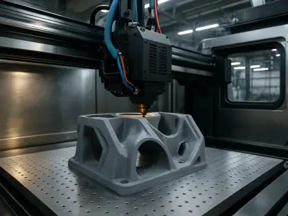
The core technology enabling this feat is known as Two-Photon Polymerization (TPP), a high-resolution 3D printing technique that utilizes a near-infrared laser to trigger chemical reactions at a specific focal point. By focusing the laser through a high-numerical-aperture objective lens, researchers can solidify a liquid photosensitive resin with a precision as fine as 100 nanometers. In this specific application, the biocompatible resin was carefully injected into the cytoplasm of the cells, where it remained in a liquid state until targeted by the laser. This localized polymerization allows for the creation of intricate three-dimensional structures without harming the surrounding organelles or the cell membrane. The specificity of the two-photon process ensures that only the material at the exact focal point hardens, leaving the rest of the cell’s internal environment untouched. This level of control is essential for building functional tools inside living systems without causing fatal trauma to the host.
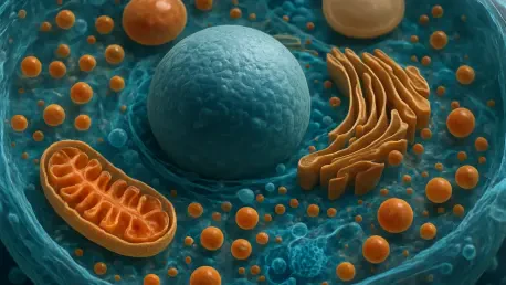
Choosing the right biological environment was just as critical as the laser technology itself, leading the research team to select HeLa cells for the initial proof-of-concept. Known for their durability and rapid division, these human cancer cells provided a robust platform to test the limits of intracellular manufacturing. The process involved a delicate balance of chemical concentration and laser intensity to ensure that the photo-initiators within the resin did not produce toxic byproducts during the solidification phase. Remarkably, the cells exhibited a high tolerance for the presence of the foreign synthetic polymers, maintaining their shape and metabolic activity throughout the procedure. This resilience indicates that the cytoplasm can serve as an effective substrate for various synthetic materials, provided the chemistry is finely tuned to avoid an immune response. The success of this methodology establishes a standardized protocol for future experiments involving even more complex biological entities and diverse material compositions.
Biological Responses and Cellular Continuity
One of the most significant findings of this research was the observation that cells containing 3D-printed structures continued to undergo normal biological processes, including cell division. Scientists monitored the HeLa cells as they progressed through the stages of mitosis, noting that the presence of a rigid synthetic object did not impede the mechanical movements required for the cell to split. In many instances, the printed structures were naturally partitioned or inherited by one of the daughter cells, which then continued to thrive as if the object were a native organelle. This persistence suggests that the cell’s internal transport systems and structural proteins can adapt to the presence of large, non-biological intrusions. The fact that the synthetic “cargo” does not trigger immediate apoptosis or cell death opens up the possibility of long-term integration. This stability is a prerequisite for any medical application that requires a synthetic component to function over an extended period.
Beyond simple geometric shapes, the ability to print microscopic barcodes directly within the cellular interior offers a transformative tool for the field of cytology and tracking. Traditional methods of labeling cells often rely on fluorescent dyes or genetic markers that can fade or mutate over time, leading to a loss of data in complex, multi-generational studies. A 3D-printed physical barcode, however, provides a permanent and unique identifier that can be read using standard microscopy techniques without the risk of degradation. This development allows researchers to track individual cell lineages in a heterogeneous population with unprecedented accuracy, observing how specific cells respond to treatments or environmental changes. This structural labeling system could revolutionize how we understand cancer metastasis or tissue regeneration by providing a reliable way to map the movement and fate of specific cells. The implications for high-throughput screening and personalized medicine are substantial.
Future Applications and Bioelectronic Integration
Advancing Toward Functional Intracellular Bioelectronics
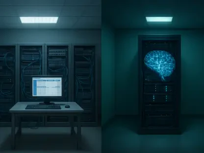
Looking ahead, the focus of this technology will likely shift from passive shapes to functional components that can interact with the cell’s internal signaling pathways. The integration of conductive polymers or responsive hydrogels could lead to the creation of intracellular sensors that monitor pH levels, calcium concentrations, or protein synthesis in real time. Unlike external sensors, these internal devices would provide a direct window into the molecular dynamics of a living system, offering data that was previously inaccessible to researchers. This approach naturally leads to the concept of “cyborg cells,” where biological functions are augmented or repaired by synthetic machinery at the scale of a single micrometer. Such advancements could allow for the correction of metabolic disorders by introducing synthetic organelles designed to perform missing enzymatic functions. This represents a new frontier where biology and electronics merge into a single, cohesive unit.
The prospect of targeted drug delivery also gains a new dimension when manufacturing can occur inside the target cell itself. Instead of relying on systemic circulation to carry medicine to a specific location, researchers could potentially print “smart” drug reservoirs that release their contents only when specific biological triggers are met. These reservoirs could be designed to degrade at a controlled rate, ensuring a steady supply of therapeutic agents exactly where they are needed most. This localized approach minimizes side effects and increases the efficacy of treatments for complex diseases like cancer or neurodegenerative conditions. As the field moves from 2026 to 2028, the refinement of these biocompatible materials will be paramount. Developing resins that are not only non-toxic but also biodegradable will ensure that these synthetic structures can be naturally cleared by the body once their mission is complete. This focus on life-cycle management is essential for clinical adoption.
Implementation and Ethical Considerations
The transition of this technology from a laboratory setting to clinical practice required a comprehensive evaluation of long-term biocompatibility and ethical implications. Researchers focused on standardizing the chemical composition of photo-sensitive resins to ensure they did not interfere with the genetic integrity of the cells over multiple generations. It was discovered that the mechanical properties of the printed objects, such as stiffness and porosity, played a crucial role in how well the cell integrated the foreign body. By mimicking the elasticity of the natural cytoskeleton, engineers produced structures that moved in harmony with the cytoplasm. These refinements were necessary to move beyond simple proof-of-concept models and toward reliable medical tools. Furthermore, the ability to print inside living tissue opened up discussions regarding the limits of biological enhancement and the potential for unintended consequences in the human germline.
The path forward for intracellular 3D printing involves the creation of multi-material structures capable of responding to external stimuli like magnetic fields or light pulses. This allowed for the remote manipulation of cell behavior, providing a non-invasive way to guide tissue growth or stimulate neural pathways. Scientists established clear safety frameworks to ensure that synthetic-biological hybrids remained under strict control, preventing any autonomous replication of the printed components. Actionable next steps for the industry included the development of portable TPP systems that could be used in clinical environments for real-time diagnostic printing. By focusing on these practical implementations, the scientific community ensured that the breakthrough at the Jožef Stefan Institute served as a foundation for a new era of molecular medicine. The successful integration of manufactured parts into living cells ultimately changed the definition of what is possible in the field of synthetic biology.