
The persistence of periprosthetic infections at the site where metal exits the skin remains one of the most significant challenges in modern prosthetic engineering. For several decades, the primary method for attaching artificial limbs has relied on the socket-based prosthesis, which requires the residual limb to fit snugly into a rigid or semi-rigid cup. While this technology has seen incremental improvements, it is fundamentally restricted by the physiological limitations of soft tissue, as skin and muscle were never designed to support the full weight of the human body under the repetitive strain of walking. This mismatch frequently leads to chronic pain, pressure sores, and tissue necrosis, which can become so debilitating that patients eventually choose to abandon their prosthetic devices entirely in favor of wheelchairs. Osseointegration has emerged as a transformative alternative, anchoring the prosthesis directly into the femur or humerus to allow for direct skeletal loading. This connection provides a phenomenon known as osseoperception, enabling patients to feel the vibration and texture of the ground through their own bones. Despite this promise, traditional solid implants have often struggled with mechanical loosening and a process called stress shielding, where the metal takes on too much load and causes the surrounding bone to weaken and wither away.
Biomimetic Engineering: The Role of Porous Lattices
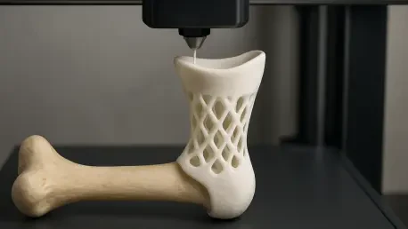
Recent breakthroughs in additive manufacturing have redefined the internal architecture of bone implants by replacing solid metal stems with intricate, 3D-printed porous structures. These lattice designs are engineered to mimic the natural trabecular geometry of human bone, which possesses a specific level of porosity and elasticity that solid materials simply cannot replicate. By utilizing advanced titanium alloys and high-precision laser powder bed fusion, engineers can now create implants that allow the patient’s own bone cells to migrate into the metal itself. This process, known as biological fixation, creates a living bond between the biological and artificial components, significantly reducing the likelihood of the implant shifting or detaching over time. Unlike traditional manufacturing, which is limited to subtractive methods like milling or turning, 3D printing allows for the creation of internal channels and graduated densities that can be tuned to match the mechanical properties of a specific patient’s cortical bone, thereby ensuring a more harmonious transition of forces across the entire interface.
The effectiveness of these porous structures is further enhanced by the ability to customize the surface chemistry and topography at a microscopic level. In the current landscape of 2026, researchers have successfully demonstrated that specifically sized pores, generally ranging between 300 and 600 micrometers, provide the optimal environment for vascularization and bone ingrowth. This architectural precision ensures that the implant is not just a foreign object sitting inside the medullary canal but becomes a structural extension of the skeleton. Furthermore, 3D printing enables the production of functionally graded materials, where the stiffness of the implant varies from its center to its outer edge. This helps to eliminate the “stress shielding” effect, as the implant can flex and compress in a manner that is similar to the host bone. By encouraging the bone to remain active and healthy through proper stimulation, these 3D-printed lattices are extending the functional lifespan of bone-anchored prosthetics, potentially allowing them to last for the remainder of a patient’s life without the need for revision surgery.
Structural Innovations: The Cloverleaf Sleeve and Dual Fixation
One of the most significant mechanical advancements in recent years is the development of modular implant configurations that move beyond a simple internal stem. Engineers have introduced the concept of an extramedullary cloverleaf sleeve, a 3D-printed component that fits over the exterior surface of the bone while an internal rod provides stability within the marrow canal. This dual-fixation strategy essentially sandwiches the remaining cortical bone between two supportive layers, which dramatically increases the surface area available for osseointegration. The cloverleaf design is particularly effective because it provides a massive boost to the structural integrity of the femur, especially in the crucial weeks following surgery when the bone is most vulnerable. By gripping the bone from both the inside and the outside, this design minimizes the tiny movements, or micro-motions, that often prevent successful healing and lead to the formation of fibrous tissue instead of solid bone.
Beyond providing a more secure anchor, the cloverleaf sleeve is specifically designed to manage the complex torsional forces that occur when a person pivots or turns while walking. In traditional bone-anchored systems, the twisting motion of a single internal stem could create enough torque to crack the bone or cause the implant to spin out of place. To solve this, the latest 3D-printed models incorporate integrated anti-rotation screws that lock the sleeve into the bone’s outer layer. These screws act as secondary stabilizers, ensuring that the prosthesis remains perfectly aligned even during high-impact activities or sudden changes in direction. This level of mechanical stability is vital for the patient’s confidence, as it eliminates the sensation of “slipping” that many early osseointegration patients reported. The combination of the internal stem and the external sleeve creates a robust mechanical bridge that can handle the three-dimensional stresses of a modern, active lifestyle, providing a level of security that was previously unattainable with older, single-component designs.
Digital Validation: Finite Element Analysis and Virtual Stress Testing
The design of these sophisticated 3D-printed implants relies heavily on the use of “digital twins,” which are highly accurate computational models of a patient’s unique anatomy. Before a single gram of titanium is printed, engineers utilize Finite Element Analysis to simulate how the proposed implant will perform under real-world conditions. By importing high-resolution CT scans into specialized software, researchers can visualize exactly how weight is distributed through the bone-implant interface during various phases of a walking cycle. These simulations can apply virtual loads ranging from 600 to 900 Newtons, representing the force of a person standing or walking, while simultaneously testing for torsional moments that occur during pivoting. This predictive modeling allows designers to identify potential “hot spots” where stress might reach dangerous levels, enabling them to refine the thickness or porosity of the implant to prevent future bone fractures or mechanical failure before the patient ever enters the operating room.
The insights gained from these digital simulations have confirmed that the new cloverleaf-sleeved designs offer a vastly superior stress distribution compared to traditional intramedullary rods. In the virtual environment, researchers observed that the sleeve effectively disperses mechanical energy across a much larger portion of the femur, preventing the concentration of force at the tip of the implant, which is a common point of failure. This improved “load sharing” ensures that the bone remains healthy and stimulated throughout its length, rather than being bypassed by a stiff metal rod. Additionally, the digital tests allow for the optimization of the 3D-printed lattice, ensuring that the porosity is high enough to encourage bone growth but low enough to maintain the required structural strength. This iterative digital process has significantly accelerated the development of bone-anchored technology, as it reduces the reliance on traditional trial-and-error methods and ensures that every manufactured implant is mathematically optimized for its specific mechanical environment.
Clinical Validation: Biological Integration and Animal Models
To bridge the gap between computer simulations and human application, researchers have turned to large-animal models that closely mirror human skeletal behavior. Small-tailed Han sheep have become a preferred model for these studies because their bone remodeling rates and weight-bearing patterns are remarkably similar to those of humans. In these preclinical trials, surgeons have implemented a technique called decortication, which involves the strategic removal of a thin layer of the bone’s outer shell to expose the nutrient-rich layers beneath. This surgical intervention triggers the body’s natural inflammatory and healing responses, essentially “tricking” the bone into weaving itself more aggressively into the 3D-printed porous lattice. By creating this intentional injury, the body’s repair mechanisms are localized exactly where the implant requires the most stability, resulting in a faster and more robust integration process.
The results from these animal trials have provided definitive proof that 3D-printed implants can survive the rigors of daily movement. Microscopic analysis of the retrieved bone samples after the trial periods showed that the sheep’s natural bone had successfully permeated the titanium lattice, creating a seamless composite material. There was no evidence of the chronic inflammation or infection that typically plagues transcutaneous implants, suggesting that the tight seal formed by the integrated bone may act as a natural barrier against bacteria. Furthermore, the implants remained stable even though the sheep were not restricted in their movements, proving that the dual-fixation design can handle the uncontrolled loading of a living subject. These findings have laid the groundwork for the next generation of human trials, providing the clinical evidence needed to move 3D-printed osseointegration from an experimental procedure to a mainstream medical solution for individuals with limb loss.
Toward a Personalized Future: The Shift to Individualized Care
The convergence of additive manufacturing and advanced imaging is moving the field of prosthetics away from “off-the-shelf” solutions and toward a future of fully personalized healthcare. Because every amputation results in a unique residual limb shape and bone density profile, a one-size-fits-all approach is inherently flawed and often leads to secondary complications. 3D printing allows clinicians to produce implants that are perfectly contoured to the patient’s remaining bone, regardless of how irregular or damaged it may be. This level of customization ensures that the implant sits in the optimal anatomical position to restore natural gait and balance. Beyond the shape, the internal stiffness of the printed metal can be tailored to match the specific bone quality of the individual, whether they are a young athlete with high bone density or an older adult with signs of osteopenia. This personalized approach reduces the risk of post-operative complications and significantly shortens the rehabilitation timeline.
The medical community prioritized these technological integrations throughout 2026 to ensure that the transition from surgery to full mobility became as seamless as possible for patients. Researchers demonstrated that the synergy between computational planning and 3D printing could eliminate many of the mechanical failures that previously defined the field of osseointegration. By moving forward with a focus on biological harmony and structural customization, the industry moved closer to a reality where artificial limbs functioned as true biological extensions of the user. Future considerations will likely involve the integration of sensors within the 3D-printed lattice to monitor bone health and implant stability in real-time. This proactive approach to post-surgical care would allow doctors to detect potential issues before they become symptomatic, ensuring that the next generation of bone-anchored prosthetics provides a lifetime of reliable, pain-free mobility. The era of the cumbersome and uncomfortable socket is rapidly being replaced by a sophisticated, skeletal-integrated architecture that honors the complexity of the human body.