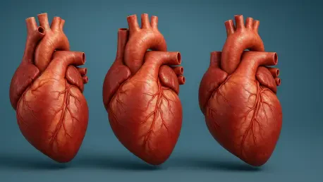

Imagine the overwhelming silence that follows a diagnosis when a cardiologist explains that a newborn’s heart, an organ barely the size of a walnut, possesses structural defects that require immediate surgical intervention. For thousands of families across Australia and beyond, this moment marks the beginning of a complex and often frightening journey characterized by medical jargon and abstract diagrams. To address this profound communication gap, a collaborative initiative known as the Heartrium project has emerged through a partnership between the University of Melbourne and the Royal Children’s Hospital. By developing the world’s first anatomically accurate, three-dimensional models of pediatric hearts, researchers are transforming how clinical information is shared between medical professionals and families. These tactile tools provide a level of clarity that traditional two-dimensional sketches simply cannot achieve, offering a physical bridge between complex pathology and human understanding for those in crisis.
Enhancing Parental Understanding through Tactile Innovation
Transforming Medical Consultations: From Sketches to Physical Reality
Traditional medical consultations often rely on hand-drawn sketches or digital diagrams to illustrate complex congenital heart defects, yet these visual aids frequently leave parents feeling confused and disconnected during critical decision-making moments. In Australia alone, approximately 2,400 children are born each year with heart conditions that require specialized care, making the need for effective communication tools a significant health priority.
The Heartrium project, led by Associate Professor Rochus Hinkel and Dr. Charles Larson, addresses this challenge by providing color-coded, 3D-printed heart models that fit comfortably in the palm of a hand. These models are based on actual medical scans, allowing parents to touch and explore the specific anomalies of a heart at the same scale as their child’s own organ. This tactile engagement serves to reduce parental anxiety by replacing abstract fear with concrete understanding, ultimately empowering families to ask informed questions.
Empowering Families: The Impact of Personal Heart Models
A compelling example of the impact of this technology is found in the experience of the Petrusev family, whose daughter, Lili, was born with hypoplastic left heart syndrome, a condition where the left side of the heart is severely underdeveloped. Her parents noted that holding a physical 3D model of a heart at a similar developmental stage provided a transformative level of insight that could not be replicated by visual presentations on a screen.
This physical representation allowed them to grasp the spatial orientation of the defect and the mechanical goals of the proposed surgeries. Inspired by the clarity they gained, they chose to have Lili’s own heart scanned to contribute to the growing Heartrium library, ensuring that future families facing similar diagnoses could benefit from the same level of anatomical transparency. By turning a personal medical challenge into a resource for others, the project fosters a community of shared knowledge that extends far beyond hospital walls.
Advancing Clinical Excellence and Global Accessibility
Technical Precision: The Role of Digital Fabrication in Education
Beyond its immediate benefits for family communication, the Heartrium project functions as a sophisticated educational resource that leverages advanced digital fabrication processes at the University’s New Experimental Technology Lab. These models are produced using high-resolution 3D printing techniques that capture the minute, intricate structures of an infant’s heart with exceptional precision and anatomical fidelity.
Dr. Charles Larson anticipates that these specialized kits will become essential components of medical training curricula for junior doctors, nurses, and surgical residents who must master the complexities of neonatal cardiology. By providing a tangible reference for rare and varied congenital conditions, the models facilitate a deeper understanding of cardiac morphology that text-based learning often fails to provide. The use of these models also improves internal communication within multidisciplinary medical teams, as surgeons can coordinate their approaches and visualize potential challenges.
Global Standards: Transitioning to Sustainable Social Ventures
The initiative thrived through substantial philanthropic support from the Royal Children’s Hospital Foundation and Finnan’s Gift, a charity that prioritized clarity and compassion for families during their most vulnerable moments. Looking toward the immediate horizon, the University of Melbourne successfully initiated the transition of Heartrium into a social venture designed to scale the technology for global distribution.
This shift aimed to commercialize the production of the models while maintaining an ethical focus on accessibility, ensuring that hospitals worldwide could adopt these tools as a new international standard for pediatric cardiac care. Medical facilities were encouraged to integrate these 3D models into their standard patient intake protocols to bridge the gap between technical diagnosis and parental comprehension. By establishing a centralized database of 3D-printable heart pathologies, the project provided a blueprint for other medical specialties to humanize complex clinical data.